
Ocean micro life in 3D
|
On June 28, 2019 the Micro Life exhibition demonstrating 3D photos of microorganisms and ocean fauna opened in the World Ocean Museum. All photos were converted to the 3D format with the help of Triaxes 3DMasterKit. 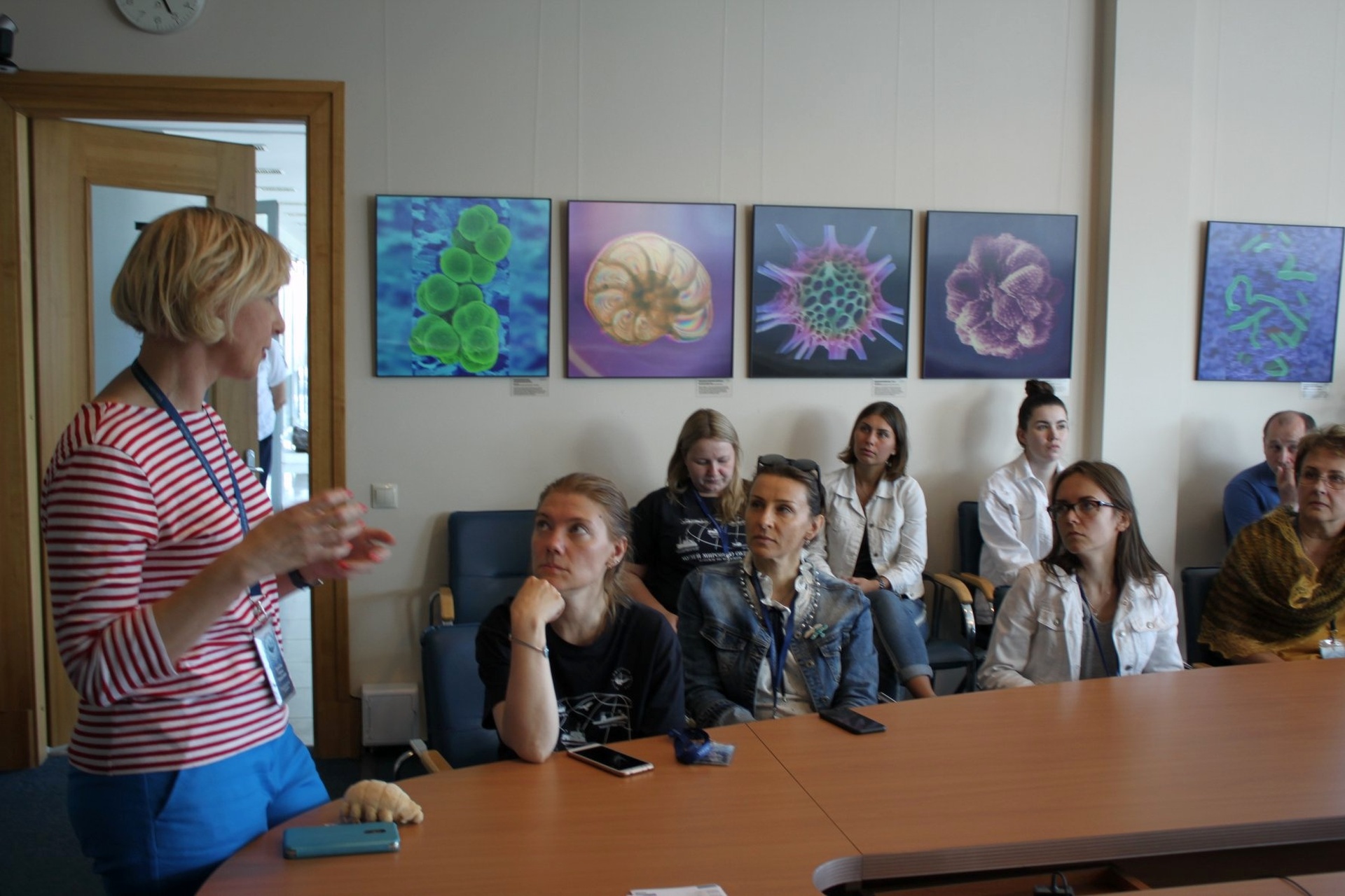 Reciting about the wonderful world of amazing microorganisms. 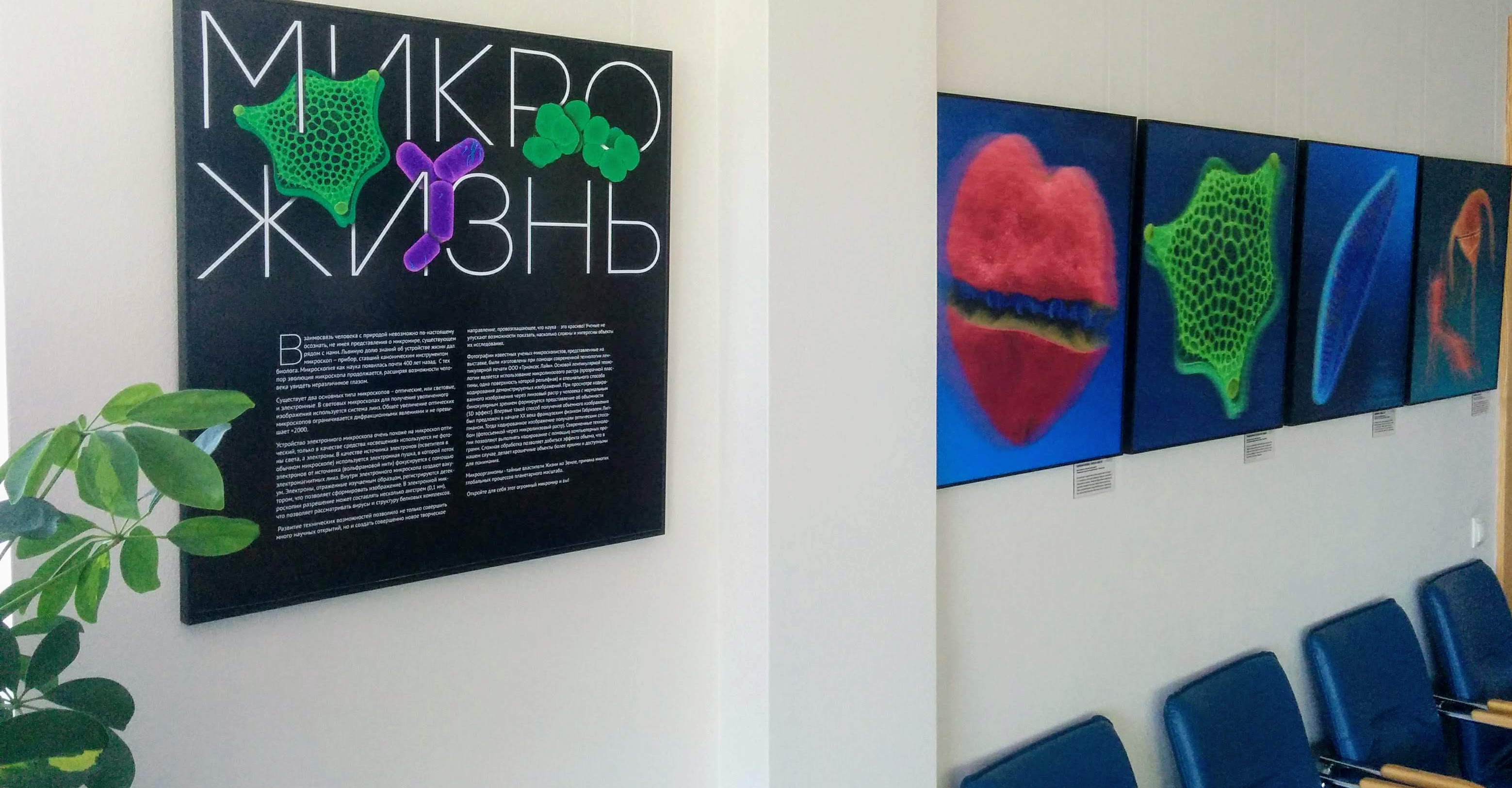 Microorganisms are secret shapers of the life on Earth. They are the reason for many global processes on the planetary scale. 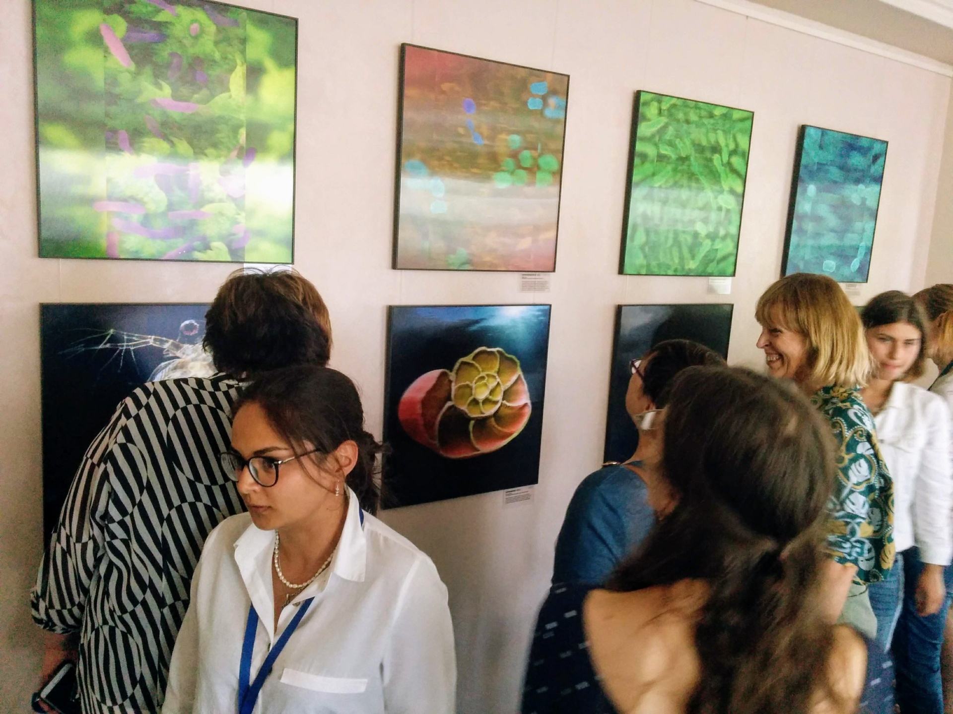 Interest and admiration of viewers are the best reward for the authors and participants of the project.  The project combining science, technology and art. Some of the pictures are demonstrated in the conference hall. The following video helps to get some understanding of how this 3D pictures look like. Their main peculiarity is that they look 3D when you watch them with both of your eyes in real time. Such an effect can be demonstrated on a regular screen im some way, and for that you must shift the camera while shooting the picture, which adds the necessary volume to the scene: background objects appear from behind foreground ones. Alexey Polyakov about the project: “As a result of interesting and intensive work, we created a series of 42 lenticular 3D images of ocean animals for the World Ocean Museum. For 2 years we had been discussing the project, creating the common concept and researching technological peculiarities. At the end of 2018 we got a chance to make it real. A selection of scientific images had to be converted from 2D to 3D, printed and delivered to the client. The December was the most intensive (every company faces the New Year rush at this time). Fortunately, all the participants of the project worked with synergy, which made it possible to get all the work done perfectly and on time. At all stages of the project cooperation with the museum staff was very constructive. With the help of Irina Baykova all the issues were speedily resolved. 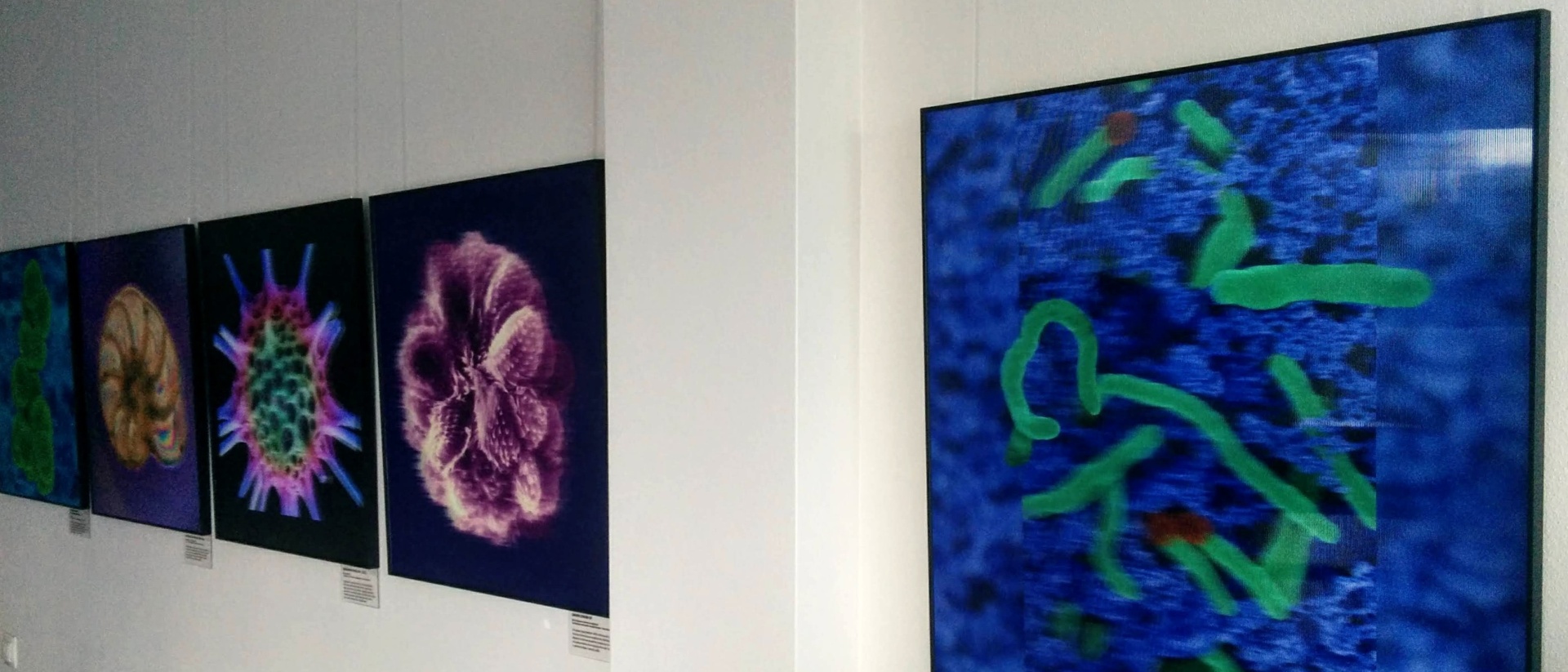 Some of the printed 3D pictures The peculiarity of the project is that the material must be, firstly, comprehensive from the scientific point of view, comply with the topic and illustrate proper biological objects (organisms); secondly, images must be attractive for the viewer from the artistic standpoint; thirdly, the peculiarities of the technology must be taken into account as objects are supposed to be in 3D. Luckily for us, we found proper photos in an agency that has a representative in Russia (it shortened the purchase process significantly). These photographs are real pieces of science and art at the same time (in case you do not know, there is an art movement in the sphere of scientific photography). Being made in 3D, the images became even more effective. Adding volume to an image is an exciting process. The work was quite intensive (we had to process 4-5 images per day). In addition, each photo has its own peculiarities. It is very inspiring, when an imagined 3D model gets its physical form and can be shared with other people”. 2D-to-3D conversion was performed with the help of modelling in 3DMasterKit developed by Triaxes. The conversion process is demonstrated in the following video. |


